Quantitative LA-ICP-MS Imaging of Elemental Distributions with Protein Correlation in 3D Tumor Models
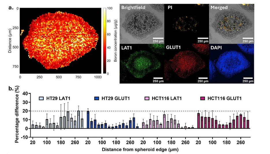
Anal. Chem. (2026) 98 (31): 23164–23178: Figure 5. Correlative analysis of elemental distribution and protein expression in tumor spheroids. (a) Representative data sets used for correlation, including a normalized boron LA-ICP-MS map, brightfield image, propidium iodide (PI) staining, and immunohistochemistry (IHC) for GLUT1 and LAT1 from an immediately adjacent section. (b) Percentage variation in protein intensity between the LA-ICP-MS section and the immediately adjacent section (±30 μm) across GLUT1 and LAT1 in HT29 and HCT116 spheroids.
This study establishes a validated workflow for quantitative LA-ICP-MS imaging of elemental distributions in 3D tumor spheroids with spatial correlation to protein expression. Optimized cryo-embedding and freeze-drying minimized analyte redistribution, while matrix-matched calibration with 31P normalization enabled reproducible pixel-level quantification consistent with bulk ICP-MS.
Applied to boron, the method achieved 10 μm spatial resolution and detection limits of approximately 7.4 ng g–1. Consecutive-section immunohistochemistry showed strong spatial agreement between elemental and protein distributions, providing a robust platform for cross-modal analysis in heterogeneous 3D biological models.
The original article
Quantitative LA-ICP-MS Imaging of Elemental Distributions with Protein Correlation in 3D Tumor Models
Fatimah Zachariah Ali*; Alexander P. Morrell; Piotr Robert Golda; Norfazlina Mohd Nawi; Premkamon Chaipanichkul; John M. McArthur; Pascal F. Durrenberger; Huda Alnufaei; Gary Royle; Kate Ricketts*
Anal. Chem. (2026) 98 (31): 23164–23178
licensed under CC-BY 4.0
Selected sections from the article follow. Formats and hyperlinks were adapted from the original.
Quantitative spatial mapping of elements in biological systems remains a methodological challenge, particularly for achieving accurate and reproducible measurements at micrometre resolution in heterogeneous 3D samples. While many imaging approaches can visualize elemental distributions, quantitative determination is limited by preparation artifacts, matrix dependent signal variability, and the lack of validated analytical workflows. (1−7) These limitations restrict mechanistic interpretation of elemental behavior in biological models and hinder comparison across studies.
Laser ablation inductively coupled plasma mass spectrometry (LA-ICP-MS) offers a powerful approach for spatially resolved elemental analysis, combining high sensitivity, broad elemental coverage, and micrometre-scale resolution. It has been widely applied to map metals, nanoparticles, and metal-based therapeutics in biological systems, including tumor spheroids, tissues, and brain models. (8−12) Despite its sensitivity and multiplexing capability, applications in complex cellular models have largely remained qualitative or relatively quantitative. (13−15) Quantitative implementation is hindered by methodological factors: sample preparation can redistribute elements, and matrix effects complicate signal interpretation, limiting reproducibility.
Here, we present a validated workflow for quantitative LA-ICP-MS imaging in heterogeneous 3D tumor spheroid models. We systematically evaluate fixation, embedding, sectioning, drying, calibration, and endogenous normalization to identify conditions that preserve spatial fidelity and analytical sensitivity. We show that endogenous phosphorus provides a robust internal normalizer, consistent with previous studies, (34−36) and demonstrate that quantitative elemental maps can be integrated with biological context through correlation with immunohistochemistry on consecutive sections. Together, this work establishes an integrated and transferable framework providing a practical platform for quantitative elemental imaging and cross-modal spatial analysis in complex 3D biological systems, with applications spanning drug delivery, nanomedicine, toxicology, and metallomics.
Experimental Section
LA-ICP-MS Instrumentation and Acquisition
Elemental imaging was performed using a 193 nm ArF* excimer laser ablation system (Iridia, Teledyne Photon Machines) equipped with a cobalt long-pulse ablation cell and coupled via an Aerosol Rapid Introduction System (ARIS) to a triple-quadrupole ICP-MS (iCAP MTX, Thermo Fisher Scientific). Instrument control and data acquisition were conducted using Qtegra software (Thermo Fisher Scientific).
Instrument performance was optimized using NIST SRM 612 glass to minimize laser-induced elemental fractionation (238U+/232Th+), maintain oxide formation below 1% (232Th16O+/232Th+), and maximize sensitivity of 59Co+, 115In+, and 238U+.
Spheroid sections mounted on glass slides were analyzed in fixed dosage raster mode with a 10 μm square laser spot, defining the spatial resolution. Imaging was performed with a laser energy density of 0.6 J cm–2, a repetition rate of 500 Hz (10 shots per pixel), and a helium carrier gas flow of 0.4 L min–1. Elemental detection was performed using Dynamic Reaction Cell (DRC) mode with oxygen as the reaction gas (0.16 mL min–1) to reduce polyatomic interferences on 31P and 32S. Two isotopes were monitored per acquisition (11B and 31P, or 11B and 32S), with dwell times of 14.3 ms (11B) and 1 ms (31P or 32S), a quadrupole total switching time of 4.7 ms, and a total acquisition time of 20 ms per pixel. A section thickness of 30 μm ensured that the imaging depth matched the physical section thickness. Slides were arranged within a four-position sample holder such that a boron-spiked fish gelatin calibration standard was analyzed within the same run to enable signal calibration.
ICP-MS Sample Preparation
For bulk quantification, spheroids were processed in parallel to LA-ICP-MS samples. Three spheroids per condition were collected into low-binding microcentrifuge tubes following incubation with BPA (4h, 37 °C). Samples were washed once with ice-cold PBS and pelleted by centrifugation (1500 rpm, 5 min). The supernatant was removed and spheroids were digested in 100 μL of 70% HNO3 (trace-metal grade, Sigma-Aldrich) at 60 °C overnight. Following complete digestion, samples were diluted in 2% HNO3 spiked with beryllium as an internal standard and analyzed using 7900 ICP-MS (Agilent). Boron concentrations were quantified against external calibration standards and expressed as total boron mass per spheroid.
Results
Overview of the LA-ICP-MS Workflow
The overall experimental pipeline is summarized in Figure 1, outlining the sequential steps from spheroid preparation to quantitative image generation. The workflow integrates cryo-embedding, sectioning, freeze-drying, laser ablation, matrix-matched calibration, signal normalization, and correlative immunohistochemistry, providing a unified framework for spatially resolved elemental quantification in 3D tumor models.
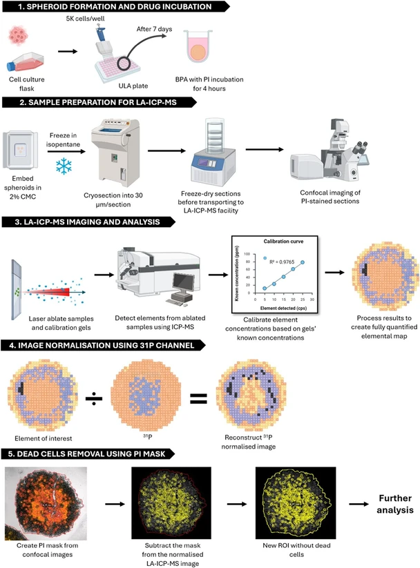 Anal. Chem. (2026) 98 (31): 23164–23178: Figure 1. Integrated workflow for quantitative LA-ICP-MS imaging in tumor spheroids. (1) Tumour spheroids are generated from cultured cells and incubated with analyte. (2) Samples are cryo-embedded in 2% carboxymethyl cellulose, frozen, sectioned, and freeze-dried prior to imaging. (3) Sections and matrix-matched calibration standards are analyzed by LA-ICP-MS to generate quantitative elemental maps. (4) Elemental images are normalized using the endogenous 31P signal to correct for variations in ablation yield and tissue density. (5) Regions corresponding to nonviable cells are excluded using a propidium iodide (PI – in red) mask derived from confocal imaging, enabling downstream quantitative analysis.
Integration of Biological Context into LA-ICP-MS Imaging
To enable spatial correlation between elemental imaging and protein expression, LA-ICP-MS maps were paired with immunohistochemical (IHC) staining acquired from immediately adjacent spheroid sections (±30 μm). Representative data sets illustrate the multimodal workflow, combining normalized boron distributions with brightfield imaging, propidium iodide (PI) staining, and IHC for GLUT1 and LAT1 (Figure 5a). PI staining was performed prior to freezing and cryosectioning to identify nonviable regions; all quantitative analyses were restricted to PI-negative regions to ensure correlation within viable cell populations.
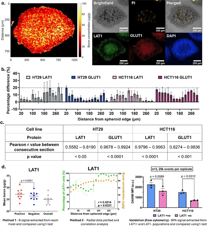 Anal. Chem. (2026) 98 (31): 23164–23178: Figure 5. Correlative analysis of elemental distribution and protein expression in tumor spheroids. (a) Representative data sets used for correlation, including a normalized boron LA-ICP-MS map, brightfield image, propidium iodide (PI) staining, and immunohistochemistry (IHC) for GLUT1 and LAT1 from an immediately adjacent section. (b) Percentage variation in protein intensity between the LA-ICP-MS section and the immediately adjacent section (±30 μm) across GLUT1 and LAT1 in HT29 and HCT116 spheroids. (c) Pearson correlation of adjacent sections (±30 μm) across GLUT1 and LAT1 in HT29 and HCT116 spheroids (n = 3). (d) Validation of finding from analysis two analysis methods (n = 3, 3 spheroids per replicate) and flow cytometric validation of BPA uptake in LAT1-positive and LAT1-negative cell populations (n = 3, no. of events = 25, 000 each replicate). Statistical significance was determined using an unpaired two-tailed t test; exact p-values are as indicated.
To assess the validity of consecutive-section correlation, protein expression was first quantified across matched sections separated by ± 30 μm. Across HT29 and HCT116 spheroids, mean variation in staining intensity between adjacent sections was 10.5 ± 6.7% and 9.5 ± 4.7% for LAT1, and 6.6 ± 0.9% and 11.4 ± 2.1% for GLUT1, respectively (Figure 5b). Overall variability across all spheroids ranged from 0.9–23.7% (n = 3 spheroids per cell line).
Coefficients of variation were 7.4 ± 2.3% and 6.8 ± 2.7% for LAT1, and 4.9 ± 1.3% and 8.3 ± 1.9% for GLUT1 in HT29 and HCT116 spheroids’ consecutive sections, respectively, indicating that spatial protein distributions were largely preserved between consecutive sections at this scale.
Consistent with this observation, Pearson correlation analysis demonstrated strong spatial agreement between adjacent sections for most proteins analyzed. GLUT1 expression showed high correlation in both HT29, (r = 0.9678–0.9924) and HCT116 spheroids (r = 0.8274–0.9836), while LAT1 expression was highly correlated in HCT116 (r = 0.9796–09963) but more variable in HT29 (r = 0.5582–0.8190) (Figure 5c). All correlations were statistically significant (p < 0.05). Section-to-section variability must be quantified before assuming that a consecutive section faithfully represents the biology of the elementally mapped section; where this condition is met, consecutive-section correlation provides a reliable means of relating elemental and protein distributions.
Having established this framework, the known relationship between LAT1 expression and boronophenylalanine (BPA) uptake was used as a biological test case. BPA is transported into cells via the large neutral amino acid transporter LAT1, providing a mechanistic link between protein expression and intracellular boron accumulation.
Region-of-interest (ROI) analysis demonstrated that LAT-1 enriched regions exhibited higher boron signal compared to LAT1-low regions in HCT116 spheroids (p = 0.0331, paired two-tailed t test; n = 3, 3 spheroids per replicate). Radial analysis further revealed a positive correlation between LAT1 expression and boron distribution (r = 0.5214, p = 0.0221, Figure 5d), supporting spatial coupling between transporter expression and uptake.
Orthogonal validation by flow cytometry confirmed that LAT1-positive cells exhibited significantly higher BPA levels than LAT1-negative cells in both cell lines (36% and 39% increase in HT29 and HCT116, respectively; p < 0.05, n = 3, 25,000 events per replicate).
Together, these results demonstrate that combined LA-ICP-MS and consecutive section IHC workflow enables biologically meaningful spatial correlation between elemental distributions and tested proteins expression in 3D tumor models.
Conclusion
This study establishes a validated workflow for quantitative LA-ICP-MS imaging in heterogeneous three-dimensional spheroid models by systematically addressing key methodological determinants of accuracy, including sample preparation, calibration, endogenous normalization, and cross-modal validation. Optimized preparation (2% CMC cryo-embedding with freeze-drying) reduced analyte loss by ∼84% relative to OCT, while matrix-matched gelatin standards enabled linear calibration (r2 > 0.99) and conversion of signal intensity to absolute concentration. Under these conditions, the workflow achieved 10 μm spatial resolution with ng g–1-level sensitivity (LOD 7.39 ± 2.24 ng g–1) and reproducible quantification across independent runs (CV < 20% across most of the spheroid radius).
Quantitative accuracy was confirmed by strong agreement with bulk ICP-MS (r2 = 0.9965; < 15% deviation above ∼3 ng), demonstrating that reconstructed LA-ICP-MS measurements provide reliable estimates of total uptake. Integration with protein expression using consecutive-section analysis was feasible when section-to-section variability was characterized (typically ∼5–12% variation; CV < 10%), enabling biologically interpretable correlations between elemental distributions and molecular markers in multicellular systems.
While volumetric reconstruction requires simplifying assumptions (e.g., radial symmetry), these do not affect the spatial correlation analyses used to interpret elemental heterogeneity. By integrating preparation optimization, validated normalization, quantitative calibration and cross-modal benchmarking within a single framework, this work moves LA-ICP-MS from relative signal mapping toward reproducible, quantitatively interpretable imaging in complex 3D biological models, providing a practical platform for mechanistic studies of drug distribution and microenvironmental heterogeneity.