Development of a Flow-through Cell for Ultrasonic Extraction (UE)─Single Particle (SP)─ICP-MS─an Approach for Nano/Microparticle Elemental and Isotopic Analysis
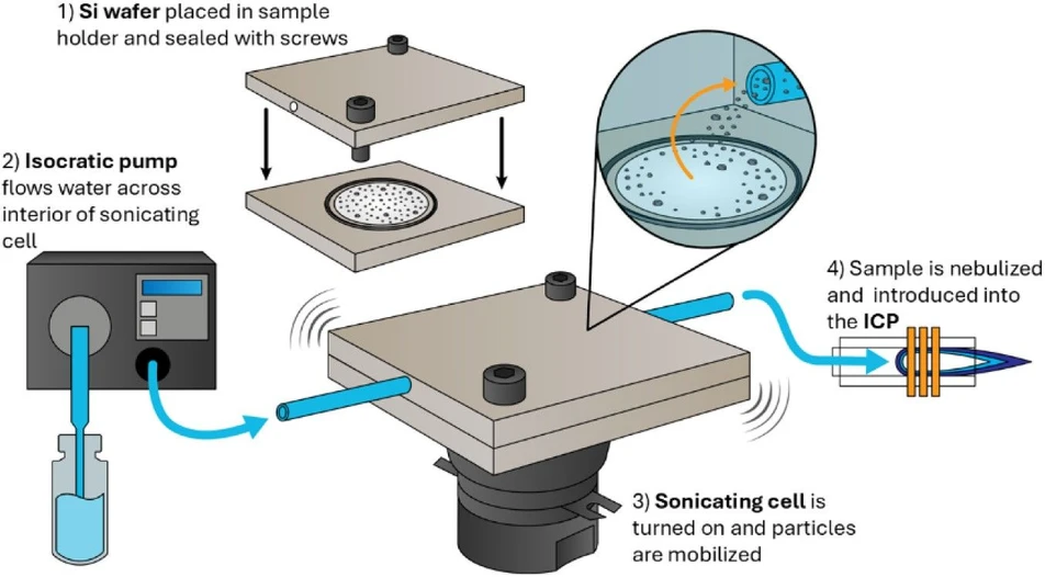
Anal. Chem. (2026): Figure 1. Depiction of the ultrasonic extraction – single particle – inductively coupled plasma–mass spectrometry approach. The sample is placed in the center of the flow through sonicating cell (1), a seal is created within the cell using a rubber O-ring and secured with fasteners. The cell is placed on top of transducer, which can be toggled on and off. An isocratic pump (2) delivers water across the sample surface, the sonication mobilizes the particles (3), and the resulting particles are nebulized and introduced into the ICP (4).
This study introduces a flow-through ultrasonic extraction cell integrated with single-particle ICP-MS for direct analysis of nano- and microparticles deposited on silicon wafers. The approach reduces sample preparation by extracting particles directly from the substrate and transferring them to the ICP-MS for elemental characterization.
Coupling the system with time-of-flight ICP-MS also enables differentiation of particles based on isotopic composition, demonstrated using isotopically tagged tungsten and nickel particles. The method offers potential for semiconductor analysis, environmental monitoring, particle synthesis, nuclear safeguards, and forensic applications.
The original article
Development of a Flow-through Cell for Ultrasonic Extraction (UE)─Single Particle (SP)─ICP-MS─an Approach for Nano/Microparticle Elemental and Isotopic Analysis
Molly K. Paul; Sarah E. Szakas; Cyril V. Thompson; Jordan S. Stanberry; Spencer M. Scott; Rachel A. Bergin; Brian W. Ticknor; Hunter B. Andrews; Benjamin T. Manard*
Anal. Chem. (2026)
https://doi.org/10.1021/acs.analchem.6c03259
licensed under CC-BY 4.0
Selected sections from the article follow. Formats and hyperlinks were adapted from the original.
Particles – specifically nanoparticles, microparticles, and colloids – have become an increasingly prominent part of analytical chemistry, with applications across diverse industries. Particles are present naturally as both biogenic (e.g., algae, fungi, proteins) and abiotic (e.g., dust, clay, wildfire aerosols) materials. Anthropogenic sources of environmental particles are often studied in forensics, and can include industrial emissions, (1) gunshot residue, (2) and even vehicle brakes. (3) The industrial applications of particle research are just as broad. Nanoparticles can be used as vaccine platforms, (4) in biocompatible polymers, (5) and for degrading environmental pollutants. (6)
Routine particle-characterization methods such as scanning electron microscopy-energy dispersive X-ray spectroscopy (SEM-EDS) (7) and dynamic light scattering (DLS) (8) are useful for imaging, elemental identification, and particle-size estimation. However, these approaches are generally low-throughput and offer limited sensitivity, particularly for elemental analysis by EDS. DLS is effective for sizing particles in samples with high particle number concentrations, while SEM-EDS – whether used alone or with automated particle analysis (APA) – supports elemental characterization but cannot scale well for high sample throughput. Importantly, neither SEM-EDS nor DLS can robustly quantify elemental or isotopic compositions at the single-particle or aggregate level.
Another approach is to dissolve the particles into solution. This method is resource-intensive and can obscure particle-level chemical variation; bulk measurements made via inductively coupled plasma–mass spectrometry (ICP-MS) collapse mixed populations into weighted averages, masking heterogeneity. This weighted average result is particularly challenging for nuclear and environmental forensics, where single particle heterogeneity is critical to assessing provenance. To meet the increasing demand for characterization of particles (i.e., elemental and isotopic composition, size, and number concentration), it is necessary to develop robust, cost-effective, high-throughput introduction systems for quantification via ICP-MS.
Single-particle (SP) ICP-MS has become a prominent technique for the determination of particle elemental compositions. (9, 10) SP─ICP-MS differs from conventional, solution-based measurements because signals are discrete rather than continuous. Measuring these discrete signals requires specialized tuning of sample introduction and detector dwell time. This technique can be coupled to a variety of mass analyzers, including quadrupole (Q), (11, 12) multicollector (MC), (13, 14) or time-of-flight (TOF). (15, 16) The quadrupole mass spectrometer is efficient at measurements of a single nuclide, while the multicollector and time-of-flight mass spectrometers specialize in measuring multiple isotopes or elements simultaneously.
Although much of the interest in SP─ICP-MS has been in the past decade, the field has a documented history of innovation. Some of the earliest studies measured Th and U particles using ICP-MS, (17, 18) and established the correlations of particle size to signal intensity and particle number concentration to signal frequency. These developments laid the foundational theory for particle counting and sizing. (19) However, translating these advancements from suspensions to particles deposited on a substrate remains limited.
Removal of particles from the substrates onto which they are deposited can be a delicate and time-intensive process, and there have been few studies which demonstrate efficient recovery. Often, particles are removed by submerging the deposition medium, usually Si wafers or C planchets, (20) in water or ethanol and sonicating. (21−23) While this method does liberate particles, it is tedious, produces dilute suspensions, and particles readily settle if not agitated frequently. Nearly all SP─ICP-MS development has been tailored to suspensions, despite the clear relevance of direct sampling for materials synthesis, semiconductors, and nuclear forensics applications and precedence of particle removal techniques used in nanofabrication (e.g., nanobubble technology, (24) aerosol mobilization, (25) laser cleaning (26)). This outpacing of need from method has, at times, produced convoluted sample introduction strategies, and, worse, loss of key population information.
To address this discrepancy, both laser-ablation sample introduction systems (27−29) and on-surface microextraction devices (30) have been pioneered. These approaches, combined with sample introduction advancements, have broadened the landscape for SP─ICP-MS. Yet a practical bottleneck remains for routine SP─ICP-MS: efficient mobilization of particles from deposition media into an ICP-MS with high sample throughput.
Described herein is a newly developed sonicating cell which mobilizes particles from Si wafers and directs them into the ICP-MS, ultrasonic extraction (UE)─SP─ICP-MS. Particles on a Si wafer surface are liberated via manually controlled in situ sonication and transported into the nebulizer via an isocratic pump with adjustable flow rates. The use of UE─SP─ICP-MS couples sonication of particles on a planchet directly in-line to an ICP-MS sample introduction, allowing for direct and rapid sampling off solid substrates with no need for sample preparation or concern of particles settling. Additionally, this sampling approach is particularly advantageous for situations where particles are likely to degrade after long periods in water or other solvents.
This device was optimized on two ICP-MS platforms (Q and TOF) using an Au nanoparticle (100 nm) standard. This in-line introduction system was further tested using synthesized, isotopically tagged W and Ni microparticles, provided by Savannah River National Laboratory in the form of particles deposited on Si wafers. (31) Analysis of particles that are fabricated in this way are especially important to the nuclear safeguards community, (32) which requires well-cataloged particle standards, and this format offers an efficient way to collect and dispatch those standards. Although there are several approaches to studying particles via ICP-MS, this method provides high extraction efficiency and allows control over the proportion of particles entering the MS while maintaining straightforward sample handling. The UE─SP─ICP-MS technique can resolve distinct isotopic populations within a single sample. This capability could support source attribution in nuclear safeguards and forensics, as well as process monitoring for particle analysis such as impurity analysis, and characterization of QC particles.
Experimental Section
Instrumentation
SEM Characterization
Samples were imaged using a scanning electron microscope (SEM; SU3800, Hitachi). The silicon wafer was mounted on a 50 mm SEM stub using copper tape, taking care to avoid disrupting particles in the center of the wafer. Secondary electron images were obtained using an accelerating voltage of 2 kV in high vacuum mode and a magnification of both 1,000× and 5,000×.
UE─SP─ICP-MS
The sonicating cell (Figure 1) was fabricated from polyetheretherketone (PEEK). It is comprised of two halves (upper and lower), which form a seal around the sample via an O-ring (Viton, Wilmington, DE). The lower half has a 1-in. diameter voided, which is recessed to hold the Si-wafer, and the other half has small prongs to provide wafer stability. Solvent (water) from the pump is introduced to the cell via a 0.025-in. diameter path and subsequently fills the internal sample volume of 660 μL, and exits the opposite side toward the ICP nebulizer. The cell is mounted on a sonicating device which is a Langevin-type transducer with a frequency of 28 kHz and a power of 60 W. The transducer is housed in a metal enclosure and coupled to a 1/4-in. aluminum plate to which the sample holder is securely fastened with 6–32 screws.
Anal. Chem. (2026): Figure 1. Depiction of the ultrasonic extraction – single particle – inductively coupled plasma–mass spectrometry approach. The sample is placed in the center of the flow through sonicating cell (1), a seal is created within the cell using a rubber O-ring and secured with fasteners. The cell is placed on top of transducer, which can be toggled on and off. An isocratic pump (2) delivers water across the sample surface, the sonication mobilizes the particles (3), and the resulting particles are nebulized and introduced into the ICP (4).
For Au nanoparticle analysis, the sonicating cell was set up in-line with an iCAP Q-ICP-MS (Thermo Scientific, Bremen, Germany). For W and Ni microparticles, the sonicating cell was set up in-line with a TOF R (TOFWERK AG, Thun, Switzerland). To measure larger, and denser, W particles, the TOF was detuned via altering the extraction voltage (voltage reduction from −161.3 V to −57.7 V). With both systems, a PFA MicroFlow nebulizer (Elemental Scientific Inc., Omaha, NE) and Cytospray spray chamber (Elemental Scientific Inc., Omaha, NE) were utilized. Type I 18.2 mΩ cm water was used as the extraction solution. The flow rate through the cell was 100 μL min–1, controlled by an isocratic liquid chromatography pump (Advion, Ithaca, NY). Sonication was continuous throughout the analysis duration. The dwell time, or integration time, for data collection from both instruments was 2 ms to ensure single particle events were captured within a few consecutive time bins.
Results and Discussion
Efficacy of Sonicating Cell ICP-MS
The sonicating cell approach was first validated using Au nanoparticle (100 nm) standards. These standards were gravimetrically prepared, placed in the cell, and sonicated continuously over a 30 min duration while mobilized particles were analyzed via Q-ICP-MS. Several measurement routines were tested, including different flow rates and sonication durations. Flow rates from 10 to 200 μL min–1 were tested, as these are commonly used in traditional ICP-MS and single particle methods. Below 100 μL min–1, inefficient particle introduction was observed while above 100 μL min–1 there were increased occurrences of multiparticle events within single time bins. It was determined that 100 μL min–1 was the preferred flow rate to resolve discrete particle signals with efficient sample throughput. This flow rate equates to approximately a 2 min sample uptake time (i.e., duration between beginning sonication and detecting particles). A time trace of the analysis interval (Figure 2) demonstrates the continuous detection of Au particles from the Si wafer. The cumulative particle count over the course of an analysis is plotted in Figure 2. The slope of this line (Figure 2, solid line) indicates that more particles are mobilized during the initial sonication (between 0 and 500 s) than at the end of the analysis duration (>800 s). The moving average particle intensity (window size 100 s) is also shown in Figure 2 (dashed line), which was used to investigate if continuous sonication directly off a surface would induce significant particle degradation or the formation of double events, as indicated by changes to the average particle intensity. The results indicate that the particles were not significantly degraded from extended sonication; the %RSDs of the average signal intensities (i.e., count per second on the quadrupole) from each 100 s window was 11%. Taking the average intensity of all particles detected (λAu = ∼1.5 × 10⁶ cps), multiparticle events (including double events, etc.) were observed at 8.6% (intensities >2 × λAu).
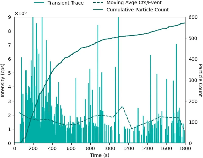 Anal. Chem. (2026): Figure 2. A time trace of signal intensity over a 30 min analysis of Au nanoparticles (100 nm). The dark green solid line follows the cumulative particle count over time (n = 571), while the dashed line traces an average particle event intensity every 100 s. Replicate experiments available in Figure S3.
Isotopic Characterization of W Particles via UE─SP─ICP-MS
Of critical importance to particle characterization is the capability to measure isotopic ratios at the single-particle scale. Because W forms semiconducting metal oxides with a range of oxidation states, it is utilized as both a primary constituent and dopant in particles, with applications in gas sensing (44) and photochromic thin films. (45) W powders (nano to micro sized) can be utilized for X-ray radiation attenuation in medical applications. (46) Furthermore, due to its high melting temperature and resistance to corrosion, W is often used to reinforce high performance materials. (47)
W particles on the Si wafers were analyzed via UE─SP─ICP-TOF-MS. Figure 3 shows SEM images of the particles prior to UE─SP─ICP-MS analysis. The resultant time-resolved data from the analysis of W particles is provided in Figure 4 and demonstrates the applicability of the UE─SP─ICP-MS method for isotopic analysis. For each particle event, multiple W isotopes (i.e., 182W, 183W, 184W, and 186W) are resolvable (Figure 4). These transient signal spikes are typical in SP─ICP-MS analysis, as individual data points are being integrated every 2 ms, allowing for ionized particle packets which have short-lived temporal durations, to be detected.
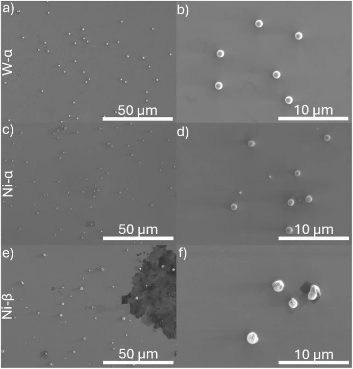 Anal. Chem. (2026): Figure 3. SEM images of particles on Si wafers. Rows correspond to sample (W, Ni-α, and Ni-β), while columns correspond to magnification (1,000× and 5,000×). Complementary postanalysis images of Ni-α are available in Figure S5. (a) is W at 1,000×, (b) is W at 5,000×, (c) is Ni-α at 1,000×, (d) is Ni-α at 5,000×, (e) is Ni-β at 1,000×, and (f) is Ni-β at 5,000×.
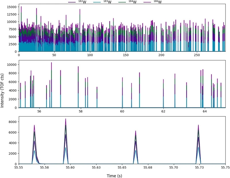 Anal. Chem. (2026): Figure 4. A time trace of the W isotopes (182W, 183W, 184W, and 186W) in the W-α particles over a 5 min analysis period. The upper panel depicts the entire analysis duration, while the lower panels depict increasingly smaller time windows.
The W particles, shown to be ∼1 μm in diameter from Figure 3a, b SEM images, are also known to have a density of 4.01 g/cm3. Due to the size and density of these particles, initial sample runs showed their signal intensities extended beyond the linear dynamic range of the TOF. To be able to detect all W isotopes of interest (182W, 183W, 186W), the extraction lens of the TOF was detuned. By dropping the extraction voltage by ∼2×, less ions were focused and extracted into the flight tube, effectively decreasing sensitivity of the measurement. To ensure the signals from the main particle population are being used to report isotope ratios, only particles with a minor isotope (183W) less than or equal to 2500 TOF counts are considered during data analysis.
Conclusions
The principal goal of this research was to demonstrate the capability of a flow through sonicating cell sample introduction system for ultrasonic extraction (UE)─SP─ICP-MS for direct sampling of solid samples for particle analysis. This approach efficiently liberates particles from Si wafers without requiring tedious sample preparation and particle preremoval into a suspension, eliminating sampling bias that can occur at these stages.
Across experiments with 100 nm Au NPs deposited onto the Si wafer, an average extraction efficiency of ∼5.3% was observed. When considering that the typical particle transport efficiency (with the employed flow rate, nebulizer, and spray chamber) is ∼5% it is possible that the UE approach achieves near full recovery of particle removal, and the limiting step is the transport of particles into the ICP. Next, the developed UE introduction was coupled to a simultaneous detection platform, ICP─TOF-MS, which allows for the isotope compositions of individual particles. The integrated approach, UE─SP─ICP-TOF-MS was employed for isotopically characterizing W and Ni isotopes within the respective particles. Briefly, the analysis of W particle was successful at determining the isotopic composition (compared to the bulk starting material used to make the particles such that the 186W/184W was within 1% relative difference (RD) from the expected value. Regarding the Ni particles, the 62Ni/61Ni was within 3% and 13% RD for the Ni-α and Ni-β, respectively.
The work presented here truly depicts a novel approach to single particle analysis, allowing for direct mobilization of particles from surfaces. Future research directions will include the following: determination of potential particle size bias derived from sonication-based liberation, quantification of particle mass, assessment of limitations in high-complexity matrices, and applicability to real-world analytical problems. The relevant spaces to apply these advancements include the semiconductor industry, nuclear safeguards and forensics, and environmental monitoring.