Electron spectroscopy for chemical analysis of liquids
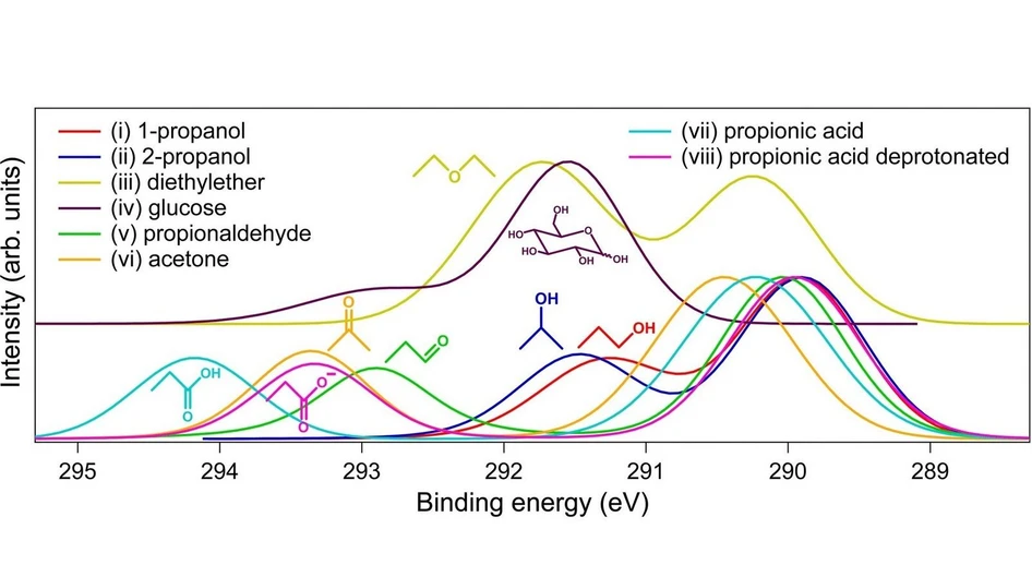
Chem. Sci., 2026,17, 6156-6164: Fig. 1 Energy-calibrated fitted C 1s photoelectron spectra of 0.5 M aqueous solutions of the compounds together with their molecular structures. Raw spectra are shown in Fig. S1 in the SI.
This study presents the first comprehensive analysis of core-level chemical shifts in aqueous solutions using Electron Spectroscopy for Chemical Analysis of Liquids (ESCAL). Absolute binding-energy calibration enabled accurate comparison of C 1s spectra across oxygenated aliphatic compounds, revealing functional-group-specific shifts strongly influenced by oxidation state, solvation, and hydration effects.
The work also resolved through-bond chemical shifts and demonstrated their dependence on molecular structure, particularly for carboxylic acid and ketone groups. These findings provide a foundation for building a predictive ESCAL reference database for liquid-phase chemical characterization and complement established structural techniques such as NMR.
The original article
Electron spectroscopy for chemical analysis of liquids
Lukáš Tomaník, Florian Trinter, Petr Slavíček and Bernd Winter
Chem. Sci., 2026,17, 6156-6164
https://doi.org/10.1039/D5SC09061J
licensed under CC-BY 4.0
Selected sections from the article follow. Formats and hyperlinks were adapted from the original.
Chemical shift is a fundamental concept in multiple spectroscopic methods, notably Nuclear Magnetic Resonance (NMR) and Electron Spectroscopy for Chemical Analysis (ESCA),1 also known as X-ray Photoelectron Spectroscopy (XPS). In ESCA, measured binding energies (BEs) of electrons reveal the elemental composition and, more importantly, chemical states of atoms. These BE changes, termed chemical shifts, ranging from a few meV to several eV, have proven invaluable for chemical analysis of solid-state surfaces and gas-phase molecules.2
The origin of ESCA chemical shifts lies in the ionization process. X-ray photons eject core-level electrons, localized on single atoms, and the recorded electron kinetic energy (KE), and hence the associated BE, reflects how strongly the electron is bound. The magnitude of the BE is governed by the net charge on the atom (initial state) and core-hole screening (final state), both of which are environment- and charge-state-dependent. Thus, any chemical modification altering charge distribution or electronic screening affects the potential field. For instance, an increased oxidation state of carbon generally increases the BE as electronegative oxygen atoms reduce electron density, increasing the positive partial charge at the carbon site, thus requiring higher photon energy for ionization. This is well-documented: gas-phase ESCA shows C 1 s BE shifts of ∼1.4 eV for alcohols and ∼3.0 eV for aldehydes/ketones compared to aliphatics,3 with similar trends in solid-state ESCA (∼1.6 eV and ∼2.9 eV for alcohols and aldehydes, respectively).4 Even secondary chemical shifts, affecting atoms one bond away, have been observed.3,4
While ESCA for solids and gases was developed in the 1960s, and low-vapor-pressure liquids were studied approximately one decade later,5–7 ESCA from volatile liquids like water became experimentally accessible only in the late 1990s due to high vacuum demands.8 Nowadays, this technique is known as liquid-jet photoelectron spectroscopy (LJ-PES).9 Additionally, combining LJ-PES with synchrotron radiation enabled precise core-level measurements and opened the door to a sharply increasing number of studies, with special emphasis on water as the most important solvent.10 Notably, recent advances in high-harmonic generation (HHG) laboratory sources promise broader access to such experiments.11,12 Despite the growing number of reported LJ-PES chemical shifts for various aqueous-phase (and non-aqueous-phase) systems, quantifying correlations between liquid-phase ESCA shifts and chemical groups or partial charges, similar to gas-phase or solid-state counterparts, has remained challenging due to the dynamic solvation environment and associated charge and energy transfers. Yet another purely experimental hurdle in LJ-PES has been accessing accurate absolute BEs associated with ill-defined potentials at the liquid-jet surface. A robust and reliable approach was introduced in 2021 by employing absolute binding-energy calibration based on low-kinetic-energy cutoffs.13 This development ensures BE comparability across different solutions and enables high precision in chemical shift measurements.14
The term ‘chemical shift’ is also central to NMR spectroscopy, which measures energy transitions of nuclear spin states in a strong magnetic field. NMR shifts arise from changes in the magnetic field caused by surrounding electrons, thus reflecting overall electron distribution.15 While the most commonly used proton (1H) NMR has a narrow chemical shift range (0–12 ppm), leading to overlapping signals in complex molecules, 13C NMR offers a much wider range (∼200 ppm), improving structural elucidation. The effect of electron distribution manifests in 13C NMR shifts of ∼10–40 ppm for aliphatic carbons, ∼55–80 ppm for alcohols, and ∼190–220 ppm for aldehydes/ketones.16
Despite sharing the terminology of ‘chemical shift,’ ESCA and NMR measure fundamentally different processes. It has been tempting to correlate the respective shifts, and these attempts have always shown significant variations.17,18 As Gelius et al.19 and Lindberg18 noted, while ESCA shifts can be interpreted via neutral-moelcule charge distributions, this is not generally true for NMR, where the dominant paramagnetic screening constant depends on excited states. Thus, a linear correlation between ESCA and NMR shifts should not be expected. Therefore, we rather view NMR and ESCA as complementary techniques for liquid-phase structural analysis.
ESCAL is enabled by experimental setups, arguably of a complexity similar to commercially available NMR spectrometers. Notably, though, the former experiments are typically performed at synchrotron radiation facilities. However, future systematic ESCAL measurements with laboratory X-ray light sources and continued improved liquid-jet delivery systems are feasible. For NMR, large libraries of documented shifts exist, helping to conveniently assign narrow peaks, measured at very high accuracy, down to hundredths of a ppm. This can be contrasted with the ∼10 meV energy resolution of ESCA from the aqueous phase, which, as we will show, is sufficient to distinguish between different molecules, charge states, and environments. We do not suggest ESCAL as a new tool for de novo structure determination of unknown compounds; rather, it provides element- and oxidation-state-specific constraints, complementing NMR for assignment and benchmarking. A unique feature of ESCA is its intrinsic ultrashort timescale (the time it takes to emit a photoelectron), thus providing, e.g., complementary information on chemical kinetics. For example, ESCA can distinguish pseudo-equivalent nitrogen atoms in an aqueous imidazole that appear equivalent in NMR.20 Moreover, ESCA can directly probe elements that are not routinely accessible by conventional NMR, such as oxygen. The two methods also differ in probing depth: NMR is generally a bulk-sensitive method, while liquid-phase ESCA primarily probes the first few molecular layers. By variation of the X-ray photon energy, one can conveniently tune between surface-sensitive and more bulk-sensitive conditions. Additionally, ESCA spectra of liquids also include unique contributions of Auger-like electrons, providing specific insight into the immediate solvation environment.21,22
This work investigates the capability of ESCA for analysis of aqueous-phase solutes, aiming to formulate general rules for liquid-phase ESCA chemical shifts and relate them to ESCA of solids/gases. We also explore the extent of secondary shifts within molecules and address the complementarity of ESCAL to NMR. We provide the first consistent study on LJ-PES chemical shifts and their origin, deriving rules and proposing general principles.
2 Results and discussion
2.1 Chemical shifts of functional groups
In this exploration of aqueous-phase ESCA, we focus on the carbon 1s electron chemical shifts in aliphatic organic compounds with oxygen-containing functional groups, a sufficient subset to demonstrate the method's capacity and lay the groundwork for future systematic extensions. The investigated functional groups include primary and secondary alcohols, ethers, hemiacetals, aldehydes, ketones, carboxylic acids, and deprotonated carboxylic acids. The molecular structures and respective photoelectron spectra are shown in Fig. 1. One observes that the peak positions differ for different compounds. The peak at ∼290 eV originates from aliphatic carbon atoms, based on experiments and calculations,23 while other peaks correspond to the functional groups. The widths of the peaks were similar for all the investigated compounds and, thus, not specific enough for their identification.
Chem. Sci., 2026,17, 6156-6164: Fig. 1 Energy-calibrated fitted C 1s photoelectron spectra of 0.5 M aqueous solutions of the compounds together with their molecular structures. Raw spectra are shown in Fig. S1 in the SI.
2.2 ESCA complementarity to NMR
Above, we discussed the fundamental and practical differences between ESCA and NMR. In the following, we discuss to what extent ESCAL may complement NMR studies, noting, however, that a direct 1 : 1 quantitative mapping of chemical shifts between the two methods is not generally expected. Yet, one can sometimes establish complementary directional trends, as we will show. We start by specifically focusing on chemical shifts in 13C NMR (Table 1, column 7) and C 1s ESCA in our set of functional groups. For better visibility, we present the data on a common axis in Fig. 3, with aliphatic carbon of isopropanol and a carboxylic acid as the low and high points, respectively. Since there is no common zero or scaling factor, we have adjusted (stretched) the NMR scale so that common trends of certain groups can be more easily identified. Overall, the comparison suggests that some groups are better distinguished in NMR while others are in ESCA. Specifically, ESCA appears to have greater resolving power for primary vs. secondary alcohol vs. ether, and for protonated (COOH) vs. deprotonated (COO−) carboxylic groups. Conversely, NMR shows a larger difference between hemiacetal vs. aldehyde, and ketone vs. deprotonated carboxylic acid. While hemiacetal and aldehyde groups exhibit very similar ESCA shifts, the respective NMR shifts are widely different and tend in different directions. Conversely, ketone and aldehyde groups show different ESCA shifts, and much higher and more similar-to-each-other NMR shifts.
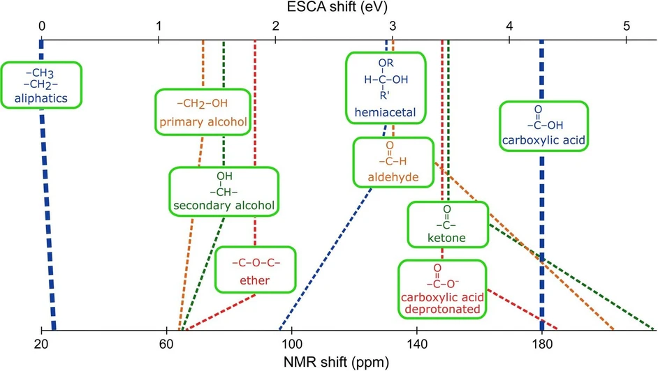 Chem. Sci., 2026,17, 6156-6164: Fig. 3 Comparison of ESCAL C 1s chemical shifts (eV) and 13C NMR chemical shifts (ppm) of various functional groups. The common scale is used for better visibility, with the aliphatic carbon of isopropanol and a carboxylic group as the low and high ends of the scale, respectively. Data for aqueous solutions of compounds summarized in Fig. 1 are used. Precise values of chemical shifts are summarized in Table 1.
3 Conclusions
We investigated whether chemical shifts in LJ-PES (liquid-phase ESCA or ESCAL) can be used similarly to ESCA of solids and gases. We recorded C 1s core-level photoelectron spectra of aqueous solutions of oxygen-containing aliphatic organic compounds and extracted absolute and accurate BEs of different carbon 1s electrons. Our results demonstrate detectable chemical shifts specific to functional groups. The trends in liquids differ from those in the gas or solid phase and highlight unique solvent effects. An observed strong correlation between experimental ESCAL shifts and calculated orbital energies enables a predictive first-order estimate of the former. ESCA and NMR chemical shifts are found to provide similar sensitivity in resolving molecular structure details, making ESCA a truly powerful alternative method to well-established NMR. The two techniques differ primarily in underlying processes, and hence observables, and time scales. Furthermore, a unique ESCA feature manifests in the capability to probe through-bond chemical shifts up to several chemical bonds away from the functional group in aqueous solutions. These secondary chemical shifts vary in strength based on functional-group type and are not universal, but dependent on specific group combinations. Observed trends enable the creation of a predictive database. This work provides the first consistent data subset, laying essential groundwork for determining empirical rules for predicting and interpreting LJ-PE spectra, similar to ESCA of gases or solids. With the expected increasing availability of liquid-phase ESCA setups, this approach can be extended to other functional groups and elements in future studies. Yet, despite its enormous sensitivity to structure determination, the performance of ESCAL experiments is currently less convenient than NMR analysis.
4 Methods
4.1 Experimental
Liquid-jet photoelectron spectra were measured at the P04 soft X-ray beamline of synchrotron PETRA III (DESY, Hamburg, Germany). We used circularly polarized light, a planar Pt-coated mirror, a laminar grating with 1200 lines/mm and 9 nm groove depth, and a vertical exit-slit size of 100 µm.34 The exact photon energy of 480.46 eV (April 2024) and 480.07 eV (December 2024) was calibrated using a precisely known liquid water valence 1b1 peak position of 50 mM NaCl aqueous solution of 11.33 eV.13 The beamline resolution with the described settings was 67 meV.
We used our EASI35 setup for the measurements. A liquid microjet under vacuum was produced using a fused-silica nozzle with an inner diameter of ∼30 µm. Solutions were delivered by a high-performance liquid chromatography (HPLC) pump at a constant flow rate of ∼1 mL min−1. A small metallic tube, placed in the main polyether ether ketone (PEEK) liquid-delivery line, enables electric grounding of the liquid or application of a defined bias voltage. The vacuum of ∼10−4 mbar in the interaction chamber was maintained using a set of scroll and turbomolecular pumps. The injected liquid was collected in liquid-nitrogen-cooled traps at the far end of the interaction chamber. The laminar-flow part of the jet, typically extending 5–10 mm from the nozzle, was irradiated by the X-ray beam in a perpendicular orientation. Photoelectrons were detected in a backward-scattering detection geometry, corresponding to an angle of 130° with respect to the light propagation direction, i.e., near magic angle, thus minimizing any differential sensitivity to the photoelectron angular distributions.35 The detection of photoelectrons was done using a hemispherical analyzer. The analyzer slit of 0.5 mm and pass energy of 50 eV correspond to the analyzer resolution of 63 meV. Thus, the total resolution as a combination of beamline and analyzer resolution is 92 meV.