A Method for the Detection of Tire Wear Microplastics in Zebrafish Guts by Laterally Resolved LA-ICP-MS-Based Elemental Fingerprinting and Chemometrics
Anal. Chem. 2026, 98, 16, 11760–11768: Graphical abstract
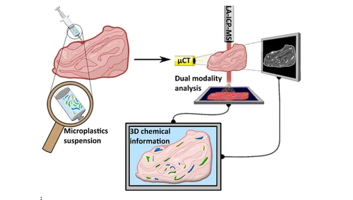
This study presents a novel method for detecting tire wear particles (TWPs) in zebrafish gut tissue using laser ablation ICP-MS, elemental fingerprinting, and machine learning. Unlike conventional FTIR or pyrolysis-GC-MS approaches, the method provides spatially resolved information and enables discrimination of TWPs from biological tissue and other particulate materials.
A random forest classification model based on multielement signatures successfully identified TWPs in complex tissue samples with a lateral resolution of 7 μm. The approach demonstrates the potential of LA-ICP-MS imaging for microplastic monitoring, ecotoxicological research, and studies of tire-derived pollution in biological systems.
The original article
A Method for the Detection of Tire Wear Microplastics in Zebrafish Guts by Laterally Resolved LA-ICP-MS-Based Elemental Fingerprinting and Chemometrics
Lukas Brunnbauer*, Šimon Juračka, Michaela Vykypělová, Lucie Vrlíková, Elisabeth Eitenberger, Pavel Pořízka, Ondrej Adamovsky, Jozef Kaiser, Gabriela Kalčíková, and Andreas Limbeck
Anal. Chem. 2026, 98, 16, 11760–11768
https://doi.org/10.1021/acs.analchem.5c07035
licensed under CC-BY 4.0
Selected sections from the article follow. Formats and hyperlinks were adapted from the original.
Tire wear particles (TWPs) are increasingly recognized as a major yet often overlooked source of microplastic pollution in terrestrial, atmospheric, and aquatic systems. (1,2) Estimations vary, but TWPs likely account for a substantial fraction of the environmental microplastics. In fact, the contribution of TWPs to total microplastic emissions in European countries has been reported to range from about 30% to over 50%. (2,3)
TWPs are generated through the mechanical abrasion of tire treads on road surfaces. (4) This wear process releases a complex mix of synthetic rubbers, fillers, and road pavement fragments. Generation of TWPs depends on several factors, including vehicle type, tire composition, road surface characteristics, and driving behavior (e.g., acceleration, braking, and cornering). (1,5−7) Notably, heavier vehicles contribute disproportionately more TWPs, and thus the support of heavier electric vehicles under the European Green Deal, despite their benefits for air emissions, may inadvertently increase TWPs. The emission of TWPs into the environment is assessed to be approximately 10% of the tire mass during use. (8) Per capita emissions are estimated to range from 0.23 to 4.7 kg per year, with a global average of 0.81 kg per year. (5)
Once released into the environment, TWPs are not well biodegradable, (9) and thus persist in soil, water, air, and sediments. (2) However, monitoring their environmental levels is considerably difficult. Conventional microplastic monitoring techniques, such as Fourier transform infrared (FTIR) microscopy, have difficulties in detecting TWPs due to the carbon black content, which absorbs infrared radiation and prevents transmission. Therefore, pyrolysis-gas chromatography–mass spectrometry (py-GC-MS) is often used to quantify TWPs in environmental and biotic samples. (10) However, this method provides only mass-based measurements, leaving unanswered the questions of particle abundance and size distribution, parameters that are essential for assessing their ecotoxicity and potential effects on human health. As a result, general monitoring campaigns often underestimate the presence of TWPs. This methodological gap underscores the need for novel techniques.
This work aims to apply laser ablation-inductively coupled plasma-mass spectrometry (LA-ICP-MS)-based imaging to identify TWPs in a biological matrix, namely, zebrafish guts, using elemental fingerprinting. Elemental fingerprinting is commonly used for classification tasks ranging from origin determination of food products (11−14) to forensic applications (15,16) using ICP-based techniques. The advantage of ICP-MS for these tasks is its outstanding sensitivity and a linear working range covering several orders of magnitude, enabling detection of major, minor, and trace constituents as well as multielement detection capabilities. This allows classification based not only on a single marker element but also on the development of more complex machine learning models to distinguish between samples based on the signals of multiple elements. When analyzing polymers with ICP-MS, only the distinct two-phase sample transport of carbon can be used to distinguish between polymer types. (17) Nevertheless, for elemental fingerprinting, the selection of elements is often based on inorganic contaminations or additives (18) present in different polymer types. (15,19) Combining ICP-MS with Laser Ablation (LA) enables laterally resolved analysis with low-μm resolution. Applying the concept of elemental fingerprinting allows us to classify each pixel of the obtained image.
In this work, we develop a method to distinguish between TWPs and the tissue of zebrafish guts and potential naturally occurring particles in the guts based on their unique elemental pattern. This is challenging due to the complex elemental fingerprint in both biological tissue and environmental TWPs since these are not well-defined matrices, and a significant variance is expected. Therefore, in a first step, we apply unsupervised clustering methods to evaluate the feasibility of distinguishing TWPs from zebrafish tissue and a range of potential occurring particles in the zebrafish gut based on a selection of detected elements. Next, we train and optimize a random forest (RF)-based classification model and evaluate its performance using independent test data. Finally, we applied the model to identify and detect TWPs in zebrafish guts.
Experimental Section
Scanning Electron Microscopy
After visual inspection of freshly cut paraffin blocks using an optical microscope (VHX-5000, Keyence, Osaka, Japan), 5 nm of gold was sputtered on the sample surface using a sputter coater (Agar Scientific, Rotherham, UK) for SEM analysis. Additionally, gold was used as a marker element in the LA-ICP-MS analysis to identify the start and end of each line scan in the transient data, facilitating synchronization with the laser log file and the following data evaluation. Surface sensitive analysis of the morphology of the samples to confirm the presence of particles in the cross sections was carried out using a scanning electron microscope (SEM, FEI Quanta 2050 FEG) in low vacuum mode and an acceleration voltage of 20 kV in backscattered electron mode (BSE).
LA-ICP-MS Analysis
LA-ICP-MS measurements were performed using an imageGEO193 laser ablation system (ESL, Bozeman, Montana, US) operating at a wavelength of 193 nm and equipped with a TwoVol3 ablation chamber. The laser ablation unit was connected to an iCAP Qc ICP-MS (ThermoFisher Scientific, Germany) via Tygon tubing (inner diameter: 1.6 mm). Ablation was conducted under a continuous helium flow of 0.8 L min–1. Argon, supplied at 1 L min–1 as a makeup gas, was combined with the sample aerosol immediately before entering the ICP using a dual concentric injector (DCI) (ESL, USA). This setup and experimental conditions result in a washout time of <100 ms. The instrument was tuned daily, maximizing 115In signal while keeping the oxide ratio 232Th16O/232Th < 1% while ablating NIST612 (Standard Reference Material, National Institute of Standards and Technology, Gaithersburg, MD).
To get an overview of elements present in TWPs that could act as marker elements, the m/z range of 6–238 was scanned using a dwell time of 10 ms per m/z. Therefore, a plateau-like signal was introduced to the ICP-MS using a line scan with a 70-μm spot size, a laser fluence of 2.4 J/cm2, a scan speed of 700 μm/s, and a repetition rate of 50 Hz. The specified m/z range was scanned 100 times and averaged to obtain representative signals.
For bulk analysis of potential naturally occurring particles and other microplastics in zebrafish guts, 20 parallel line scans (length of 5 mm) were ablated for each of the investigated materials (road dust, mussel’s tissue, lake sediment, river sediment, paraffin, gut tissue, hatched Artemia, Hikari feed, PVC, PET, PP, HDPE, and TWPs). Using a spot size of 70 μm, a laser fluence of 2.4 J/cm2, a scan speed of 700 μm/s, and a repetition rate of 50 Hz results in an ablation time of around 7 s per line. These settings resulted in a continuous representative signal for each sample. For each line scan, the ICP-MS signal was averaged and used for data evaluation.
Results and Discussion
Elemental Fingerprint of TWPs
In the first step, the elemental fingerprint, which consists of marker elements for the TWPs, was investigated. Therefore, TWPs mounted in paraffin were continuously analyzed with a large spot size (70 μm) to sample the particles as representatively as possible. Recording the mass spectrum from m/z 6–238 allowed us to identify potential marker elements in the TWPs. The mass spectrum was compared to the mass spectra of pure paraffin and a gas blank to identify potential marker elements. A wide range of environmental elements, such as C, Na, Mg, K, Ca, Cu, Fe, and Zn, were detected in the TWPs. Additionally, elements of potential anthropogenic origin, such as Ti, Mo, Sb, Nd, and Pb, were detected. Isotopic patterns of each element were verified against their natural abundances to ensure accurate identification and avoid interference-related misinterpretation.
Most biogenic elements are expected to be abundant in zebrafish tissue, as well, and thus unsuitable for distinguishing TWPs from tissue. Consequently, Zn, Ti, Mo, Sb, Nd, and Pb were selected for elemental fingerprinting. Even though Zn is a biogenic element, it was selected because it is one of the main additives (as ZnO and Zn stearate) in tires and is typically present at the g/kg level. Pb is known to occur as a contaminant in ZnO and can, therefore, be found in TWPs as a trace constituent (mg/kg). Ti serves as a catalyst in rubber manufacturing and can also be present as an additive. (29) Sb and Mo are commonly found in commercial brake pads and have been detected in several studies in road dust and TWPs. (30−32) Nd-based Ziegler–Natta catalysts are employed in the synthesis of diene-based elastomers for tire applications. (33)
To assess the feasibility of elemental fingerprinting to identify TWPs in zebrafish guts and reliably distinguish them from naturally occurring particles, in a first step, bulk analysis is carried out. Therefore, the elemental fingerprint of TWPs was compared to 12 other compounds (lake sediment, river sediment, road dust, mussel tissue, Hikari feed, and dried artemia used to feed the fish) as well as a blank zebrafish gut and paraffin. Additionally, other microplastics (PVC, PET, PP, and HDPE) were analyzed. For each sample, 20 line-scan measurements were carried out, ICP-MS signals were averaged, and data were standardized for the following unsupervised data evaluation. In a first step, principal component analysis (PCA) was calculated, and biplots for PC1 vs PC2 (Figure 1a) and PC2 vs PC3 (Figure 1b) are shown. The detected elements were the input variables. PCA enables visualization of the 6-dimensional feature space via projections and enables interpretability based on the loadings. Clear differences between the analyzed samples can be observed based on the selected elements. PET stands out with a high signal for Sb due to a common Sb-based catalyst used in PET manufacturing. (34) Lake sediment stands out with high signals for Ti, whereas river sediment shows high signals for Nd. Road dust shows significant signals for Pb and Mo, and as expected from the additives, TWP shows high amounts of Zn. In the biplots, TWPs are well separated from the other compounds. To provide further insights into the elemental patterns of the different materials, a standardized (column-wise) heatmap of the observed signals is provided in the Supporting Information (Figure S3).
 Anal. Chem. 2026, 98, 16, 11760–11768: Figure 1. PCA for the bulk analysis of potential (naturally) occurring particles/organics in zebrafish guts and TWPs (a) shows the biplot for PC1 vs PC2, (b) shows the biplot for PC2 vs PC3.
Application to Detect TWPs in the Zebrafish Gut
In the previous section, we demonstrated that the model can reliably distinguish between particles and tissue based on LA-ICP-MS elemental images. In the final step, the model was applied to identify TWPs directly in the zebrafish gut. Therefore, we applied the developed model to evaluate elemental maps obtained from zebrafish guts with inserted TWPs (elemental maps for the two analyzed samples are shown in the Supporting Information in Figures S8 and S9). Classification results of the developed model are listed in Figure 6. For the sample TWPs in gut 1, several particles in the center of the sample are identified as TWPs, revealing not only the position but also the shape of the individual particles, which matches the shape of the particles observed in the SEM pictures. In the section of the analyzed gut, no other particles were visible. For the sample TWPs in gut 2, several TWPs were detected using the developed method. Aligning the results with the SEM pictures enabled correlating the classification results with individual particles observed in the SEM pictures. For this sample, similar to the blank samples, many particles were found in the zebrafish gut in the SEM pictures. Combining SEM pictures with LA-ICP-MS-based elemental fingerprinting enabled clear identification of TWPs among the particles found in zebrafish guts. Additionally, it should be noted that the information depth of SEM pictures and LA-ICP-MS analysis is different. While SEM analysis is expected to have an information depth showing only particles in the first 2–4 μm of the sample, LA-ICP-MS analysis ablates 8 μm. Therefore, TWPs that were not fully observed in the SEM picture due to still being (partially) covered by paraffin may be detected by the developed LA-ICP-MS method. Additionally, it should be highlighted that the obtained pixel size for LA-ICP-MS images corresponds to 7 μm. To estimate the smallest detectable particle size, we apply geometric considerations and not a classical LOD approach based on a mass fraction. A classical LOD approach is not feasible in our scenario, since the developed classifier is trained on pure pixels that are fully contained within an individual TWP. Therefore, even if we were able to analytically detect the signal of the marker elements for a < 7 μm particle within one pixel, the classifier would not reliably identify the pixel as TWP since the absolute signals would be lower. Therefore, only a particle that produces at least one pixel fully contained within the particle is detectable. Accordingly, based on geometric assumptions and under idealized conditions, the smallest detectable particle size is estimated to be 14 μm, corresponding to twice the laser spot size. This requirement ensures that at least one laser shot is fully contained within the particle, independent of raster alignment. Consequently, 14 μm represents the theoretical lower limit of detectable particle size for the developed method. It should be noted that the practical detection limit may be influenced by additional factors such as particle shape/morphology and beam profile.
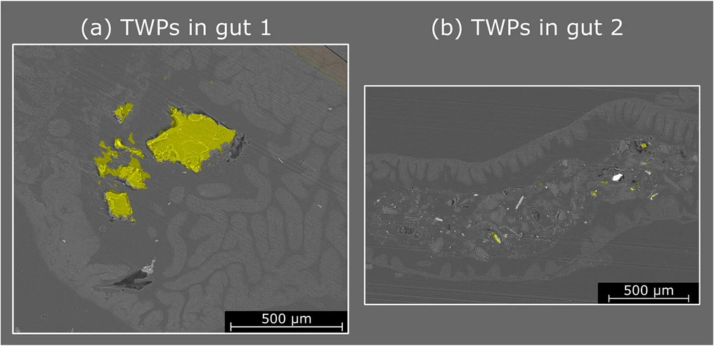 Anal. Chem. 2026, 98, 16, 11760–11768: Figure 6. Classification results of images obtained from two different zebrafish guts exposed to TWPs overlaid with SEM pictures (a, b). Pixels classified as TWPS are marked in yellow. Pixels classified as tissue or paraffin are transparent. The area analyzed by LA-ICP-MS was larger than the SEM picture; therefore, the area is not marked.
Conclusion
Despite their widespread occurrence, the detection of TWPs remains challenging due to their complex and heterogeneous composition, which differs fundamentally from widely studied thermoplastics and thermoset microplastics. Here, we demonstrate that LA-ICP-MS elemental imaging combined with random forest classification provides a powerful approach for detecting TWPs in biotic tissue. While individual marker elements such as Zn or Ti were insufficient to identify TWPs due to overlapping with signals from biological tissue, the multivariate classification model successfully achieved reliable discrimination based on combining multiple marker elements. The developed model showed excellent performance on independent test data, minimizing false positives and accurately identifying TWPs even in the presence of natural particles. Application to zebrafish gut samples confirmed that TWPs could be detected. These findings highlight the potential of elemental fingerprinting combined with machine learning to overcome the limitations of existing analytical methods for TWP monitoring. Beyond advancing microplastic research, this approach provides a framework for studying particle uptake, translocation, and distribution in organisms and humans, contributing to a better understanding of the ecological and health implications of TWPs. The growing regulatory and environmental focus on tire wear emissions underscores the urgent need for advanced analytical methods capable of identifying TWPs with high accuracy in complex matrices. Future instrumental improvements resulting in increased sensitivity may further boost the lateral resolution of the developed method. ICP-TOF-MS-based detection, enabling simultaneous detection of a wide m/z range, could further improve the classification approach’s robustness and extend the approach to other biological matrices and particles.