Photocatalytic degradation and transformation of pharmaceuticals using exfoliated metal-free g-C3N4
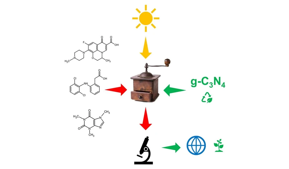
iScience. 2025 Oct 30;28(12):113899: Graphical abstract
This study evaluates thermally exfoliated metal-free graphitic carbon nitride (g-C₃N₄) as a visible-light photocatalyst for removing pharmaceutical micropollutants, including ofloxacin, diclofenac, and caffeine. The material exfoliated for two hours showed the highest degradation efficiency, achieving more than 95% removal of ofloxacin and diclofenac and around 80% removal of caffeine within 120 minutes.
Liquid chromatography coupled with high-resolution tandem mass spectrometry (LC-HRMS/MS) was used to identify degradation products and elucidate transformation pathways. The results demonstrate the potential of exfoliated g-C₃N₄ for efficient photocatalytic water treatment while providing detailed insight into the fate of pharmaceutical contaminants.
The original article
Photocatalytic degradation and transformation of pharmaceuticals using exfoliated metal-free g-C3N4
Petr Praus, Anna Gavlová, Jan Hrbáč, Kristina Schmidtová, Petr Bednář
iScience. 2025 Oct 30;28(12):113899
licensed under CC-BY 4.0
Selected sections from the article follow. Formats and hyperlinks were adapted from the original.
Pharmaceuticals of both anthropogenic and natural origins contribute to the growing issue of micropollutant contamination worldwide. These substances can enter aquatic systems through various means, including the excretion of non-metabolized drugs by humans and animals, improper disposal of unused medications, and industrial and agricultural runoff. Urban wastewater treatment plants are significant point sources, as they often fail to completely remove these compounds during treatment processes. Pharmaceuticals have been found in surface water, groundwater, soil, and sediments. In addition to the complexity of these samples, they often exist in trace concentrations at the level of nanogrammes or micrograms per liter or kilogram in liquid or solid samples of aquatic systems, which makes their analytical determination difficult.
Traditional wastewater treatment based on the activated sludge process is only partially effective in removing pharmaceuticals.1,2 In principle, adsorption and separation processes retain pharmaceuticals; however, they do not address the ultimate disposal of the separated organic compounds. In contrast, photocatalysis enables the removal of pharmaceuticals from aqueous environments by their degradation.3 Photocatalysis is part of advanced oxidation processes (AOPs) that can be used to treat organic pollutants in water. AOPs are usually performed using ozone, hydrogen peroxide, persulphate, peroxymonosulfate, ozone, sonolysis, and iron salts in the Fenton process.4 Photocatalysis has also been used for various reactions, such as the reduction of CO2,5 degradation of dyes6 and other organic compounds,7 hydrogen evolution by water splitting8 or adding NaBH4,9 fixation of nitrogen,10 reduction of Cr(VI),11 synthesis of hydrogen peroxide,12 water disinfection,13 and gene removal.14 The application of Fe-MOF-based composites is also a promising strategy for the photocatalytic removal of environmental pollutants.15
Most photocatalytic applications are based on TiO2, but graphitic carbon nitride has also been found to be a suitable photocatalyst for the degradation of various organic compounds, including pharmaceuticals. This material has been investigated over the last decade because it can be activated by visible irradiation owing to its narrow band gap (2.7 eV) and diamond-like physicochemical properties, such as mechanical, thermal, and chemical stability.16 The popularity of g-C3N4 also lies in its simple preparation via thermal synthesis from nitrogen-rich organic precursors, such as cyanamide, dicyandiamide, and melamine, in relatively high quantities at a low cost.
However, the low specific surface area and fast recombination of photoinduced electron - hole pairs, resulting in low quantum efficiency, are drawbacks of g-C3N4. The low specific surface area of pre-prepared (bulk) g-C3N4 can be increased by exfoliation using various methods, such as chemical oxidation with K2Cr2O7, concentrated sulfuric acid, organic liquids, sonication, ion intercalation, and thermal exfoliation. Thermal exfoliation, notable for its simplicity, ease of implementation, and environmental friendliness, is often used. In addition, the physico-chemical properties of g-C3N4 can be fine-tuned by coupling with anions, metal cations, metal nanoparticles such as Au and Au/Pd, Ag, Cu, Cu/Co, or semiconductor particles such as TiO2, ZnO, FeS2, Al2O3, SnO2, WO3, Ag3VO4, BiVO4, BiOBr, BiIO4, and Cu3V2O8. Another approach is doping with metal and non-metal elements, such as O, S, and P.
This study aimed to investigate the photocatalytic degradation of pharmaceuticals, including their transformation products. For this purpose, bulk and thermally exfoliated g-C3N4 samples were used in this study. This is a simple, one-component, metal-free, and low-cost photocatalyst with potential application in water treatment technology. Moreover, its photocatalytic stability during the degradation of organic substances has been reported in the literature.17 The photocatalysts were synthesized from dicyandiamide and characterized by elemental analysis, electron microscopy, X-ray diffraction, physisorption of nitrogen, and infrared, UV-Vis, photoluminescence, and electron paramagnetic resonance spectroscopies. In photocatalytic experiments, the common and widespread pharmaceuticals ofloxacin, diclofenac, and caffeine, selected as model compounds, were degraded under visible light irradiation at 420 nm. The degradation rates and efficiencies were evaluated, and degradation pathways were suggested based on the identification of intermediary degradation products using liquid chromatography combined with high-resolution tandem mass spectrometry.
The novelty of this study lies in the investigation of the transformation pathways of selected pharmaceuticals, such as ofloxacin, diclofenac, and caffeine. Such transformation investigations are important for possible water treatment applications, but are often omitted from reported studies. An important feature is that pure g-C3N4 can be an effective and suitable photocatalyst, which can be simply tuned through thermal exfoliation to achieve results comparable to those of other complex multi-component photocatalytic systems. Moreover, unlike other photocatalysts, such as TiO2, g-C3N4 operates under visible light irradiation.
Method details
Elemental analysis
The C, H, and N contents in the g-C3N4 materials were determined using a Flash 2000 elemental analyser (ThermoFisher Scientific, Waltham, MA, USA). The oxygen content was estimated as the remaining fraction after subtracting the measured C, H, and N content from 100%.
Scanning and transmission electron microscopy
Scanning electron microscopy and energy-dispersive X-ray spectroscopy (EDS) analyses of the g-C3N4 materials were performed using a Tescan Vega microscope (Brno, Czech Republic) equipped with a tungsten cathode. SEM micrographs were obtained using secondary electron (SE) and backscattered electron (BSE) modes with an acceleration voltage of 30 keV. The samples were gold-sputtered before analysis to ensure adequate electron conductivity.
Transmission electron microscopy was performed using a JEOL 2100 microscope equipped with a LaB6 electron gun. An accelerating voltage of 200 kV was applied. TEM micrographs were recorded using a Tengra camera (EMSIS). For the TEM analysis, the samples were dispersed in ethanol and sonicated for 5 min. One drop of this solution was placed on a copper grid with a holey carbon film and dried at room temperature.
Fourier transform infrared spectroscopy
Fourier transform infrared spectroscopy was performed using a Nicolet iS50 device (Thermo Scientific, Waltham, MA, USA) with the KBr pellet technique. The powder sample was mixed and homogenised with KBr (approximately 200 mg) and pressed at a pressure of 20 MPa to obtain a transmission pellet. The prepared pellet was placed in the holder of a transmission attachment, and the FTIR spectra were collected in the wavenumber range of 500–4000 cm−1 with a resolution of 2 cm−1. Each spectrum consisted of at least 64 scans, each lasting 1 s. Before each measurement, the background was collected to eliminate the effects of the apparatus and environment.
Results and discussion
The g-C3N4 bulk and exfoliated materials were synthesized from dicyandiamide in air, and their physico-chemical properties were extensively characterized. The photocatalytic properties were studied by the degradation of selected pharmaceuticals (ofloxacin, diclofenac, and caffeine), and the degradation products were analyzed by liquid chromatography. Transformation pathways were proposed for all three pharmaceuticals studied.
Elemental analysis
The synthesized bulk and exfoliated g-C3N4 samples were analyzed to determine their elemental compositions (Table 1). The C, H, and N contents were determined directly, while the O content was calculated by difference to reach a total of 100%. The C and N contents were similar in the exfoliated g-C3N4 samples but were generally lower than those in bulk g-C3N4 because these elements were released during the exfoliation process, indicating the formation of both nitrogen and carbon defects (vacancies). The oxygen and hydrogen contents were higher in the exfoliated materials owing to their partial oxidation in air and the formation of -OH groups, as shown in the Fourier transform infrared (FTIR) spectra (Figure 4).
Characterization by electron microscopy
The morphologies of the g-C3N4 materials were investigated by scanning electron microscopy (SEM). Micrographs of the Bulk and TEX3 samples (samples with the lowest and highest surface areas, respectively) are presented in Figure 1. Their morphologies were very similar, that is, large particles composed of aggregated flakes. The results of the SEM-EDS analysis are summarized in Table S1. The nitrogen, carbon, and oxygen contents of g-C3N4, as well as the C/N ratios, do not correspond with the results of the elemental analysis given in Table 1. The EDS results are not realistic for several reasons, namely a small amount of the analyzed materials, the impossibility to determine hydrogen, and a low sensitivity of EDS to light elements (C, N). The data can also be distorted due to the use of carbon tape to affix the powder samples. Therefore, we consider the results of conventional elemental analysis, which uses at least 0.5 g of each sample, to be reliable and realistic. However, a useful piece of information from the SEM-EDS is that the synthesized g-C3N4 materials were not contaminated by other elements.
 iScience. 2025 Oct 30;28(12):113899: Figure 1 - SEM (BSE+SE) micrographs of (A) Bulk and (B) TEX3 graphitic carbon nitride. TEM micrographs of (C) Bulk and (D) TEX3 graphitic carbon nitride. SEM analysis: the acceleration voltage was 30 keV. The samples were gold-sputtered before analysis to ensure adequate electron conductivity. TEM analysis: an accelerating voltage of 200 kV was applied. Ethanol dispersions were placed on copper grids with holey carbon films.
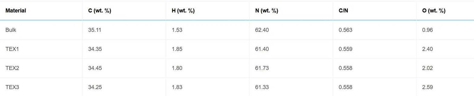 iScience. 2025 Oct 30;28(12):113899: Table 1 - Elemental composition of bulk and exfoliated g-C3N4
The transmission electron microscopy (TEM) micrographs are shown in Figure 1. The presence of worm-like structures is typical of exfoliated g-C3N4 materials. One can also see large flat flakes with different thicknesses and agglomerations. The worm-like structures were probably formed by the wrapping and deformation of flat flakes due to prolonged exposure to high temperatures.
Characterization by X-Ray diffraction
The structures of the synthesized bulk and exfoliated g-C3N4 were studied using X-ray diffraction (XRD) (Figure 2). Two typical main diffraction peaks, (002) and (100), were observed (JCPDS 87–1526, supplemental information). The (002) and (100) diffractions can be ascribed to the interlayer stacking of the g-C3N4 planes and the in-plane ordering of the nitrogen-linked heptazine units, respectively. The selected characteristics of the diffraction peaks are listed in Table 2.
The d(002) spacing of bulk g-C3N4 decreased slightly after exfoliation, while the crystallite size L(002) increased. These structural changes can be explained by the formation of expanded crystallites owing to the thermal treatment of bulk g-C3N4.
 iScience. 2025 Oct 30;28(12):113899: Figure 2 - XRD patterns of bulk and exfoliated g-C3N4 using a Cu tube operated at 40 kV and 30 mA.
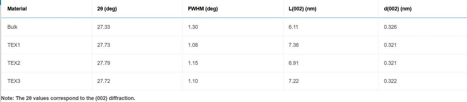 iScience. 2025 Oct 30;28(12):113899: Table 2 - XRD characteristics of bulk and exfoliated g-C3N4.
Transformation pathway of ofloxacin
Thirteen TPs were identified for ofloxacin (Table S4). Three structures corresponding to m/z 364.1278 and three structures corresponding to m/z 350.1143 are labeled with numbers 7 and 5, to which small letters (a, b, c) are added, respectively. We were able to propose four transformation pathways, which included 11 out of 13 identified TPs (TP3-TP9, see Scheme 1); TP 1 and TP 2 did not directly fit any of the pathways, so they are listed separately (Table S4). During the photocatalytic degradation, ofloxacin lost the carboxyl group, and the methylpiperazine ring was cleaved to varying degrees without further oxidation, resulting in the formation of TP 1 and TP 2. The mechanism leading to these TPs appears to be fragmentation rather than oxidation, and these TPs have not been previously documented in the literature.
 iScience. 2025 Oct 30;28(12):113899: Scheme 1 Proposed transformation pathways for the photocatalytic degradation of ofloxacin
Conclusion
Bulk graphitic carbon nitride was synthesized via dicyandiamide thermolysis at 550 °C. Exfoliated materials were prepared by the further calcination of bulk g-C3N4 at 500 °C for 1–3 h (TEX1-3). The prepared materials were characterized by elemental analysis, SEM and TEM imaging, XRD, physisorption of nitrogen, XPS, FTIR, UV-Vis, photoluminescence, and EPR spectroscopy.
The exfoliation of g-C3N4 provided a higher specific surface area, leading to more reaction sites and longer lifetimes of the photoinduced electrons and holes for the photocatalytic reactions. However, excessive thermal exfoliation led to the degradation of graphitic carbon nitride by the formation of more defects (TEX3), as documented by structural, textural, and spectroscopic analyses. Under the specific conditions of this study, the optimum exfoliation time was determined to be 2 h. The corresponding material provided a relatively high SBET, lifetime, and the highest PL intensity, which was related to the highest number of electrons and holes. It was the most active photocatalyst in this study, as confirmed by the photocatalytic degradation of ofloxacin, diclofenac, and caffeine under irradiation at 420 nm.
The superoxide anion radical was identified as the primary reactive oxygen species mediating degradation using EPR spectroscopy. The degradation rate decreased in the order of ofloxacin > diclofenac > caffeine. After 120 min, more than 95% of ofloxacin and diclofenac and approximately 80% of caffeine were degraded. Although most of the pharmaceuticals were removed, photocatalytic investigations were further focused on identifying intermediary degradation products using HPLC/HRTMS. Based on the identified intermediates, transformation pathways were proposed.
This study confirms that exfoliated g-C3N4 is a suitable metal-free, low-cost, and easy-to-prepare photocatalyst that can be activated by visible light and has potential in green technology water treatment. This also demonstrates that not only the degradation rate of the original organic compounds but also their degradation products need to be investigated. Despite significant progress, a knowledge gap remains regarding the degradation products generated by photocatalysis and, more broadly, advanced oxidation processes (AOPs). Sensitive analytical techniques are essential for the accurate identification and quantification of these transformation products.