TU Wien launches a research platform for 3D testing of bio-samples

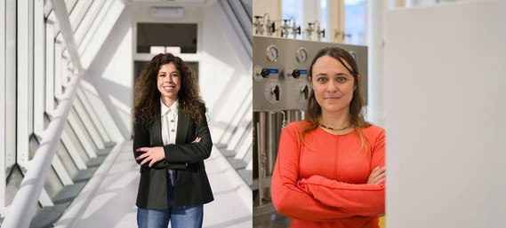
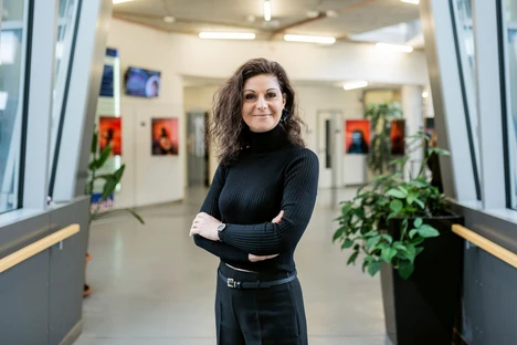
- Photo: TU Wien: Mina Petrovic during the opening event.
From individual cells and small tissue samples to fruit flies: if you want to research biological material, you often need very special equipment. The "LifeScope3D" platform has now been set up at TU Wien to provide optimal instrumentation for studying 3D multicellular structures, such as organoids and spheroids. A whole range of specialized equipment is now available at the Getreidemarkt campus, which enables characterization on various scales- starting from entire objects down to single cell or molecule analysis. However, the platform is not only interesting for biological samples, but also for other similar-sized structures, such as those produced by a high-resolution 3D printer.
Molecular biologist, Mina Petrovic, PhD, is the project manager of the new platform. LifeScope3D is the result of a collaboration between Prof. Aleksandr Ovsianikov (Institute of Materials Science and Technology), Prof. Philipp Thurner (Institute of Lightweight Structures and Structural Biomechanics) and Prof. Gerhard Schütz (Institute of Applied Physics). However, the platform will also be accessible to all other research groups - both within and outside TU Wien. LifeScope3D is intended to create a hub that promotes multidisciplinary collaborations in the field of cellular assemblies and tissue engineering. This was made possible by funding from the Austrian Research Promotion Agency FFG.
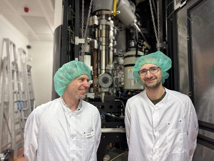
 TU Wien: The biosorter in the new lab at TU Wien.
Different measuring devices
"We can not only image biological structures, but also fully analyze them in three dimensions," explains Mina Petrovic. "This is of great importance for many applications in biology." For example, a special microscope on the new research platform can be used to illuminate the samples with so-called "lattice light sheets". Instead of a beam of light that scans the sample point by point, an extremely thin, two-dimensional light sheet is generated that penetrates the sample layer by layer. Each time, only dyes located within this thin light sheet are detected and a three-dimensional image with high contrast can then be put together on the computer.
However, optical methods are not the only way to examine samples - mechanical experiments also play an important role. The sample can be subjected to a precisely defined mechanical load. While the sample is compressed, the sample deformation is measured. Valuable findings can be derived from this, as such experiments mimic the pressures that cells have to endure in different parts of the body.
A bio-sorter is also used in the new research platform: "Biological samples are rarely uniform, but often come in a mixture of different sizes, showing different features”, explains Mina Petrovic. "In the bio-sorter, a mixture of, for example, multicellular spheroid structures is guided past a laser beam where each individual structure can be precisely analyzed and categorized at a very high speed."
 TU Wien: Computer controlled experiments with the lattice light sheet.
Accessible for everybody
Numerous research groups from different faculties at TU Wien are conducting research on biological samples - LifeScope3D will open up new analysis methods for all of them. However, the aim is also to collaborate with research teams outside of TU Wien: "In principle, LifeScope3D is open to all groups with suitable research projects," says Mina Petrovic. "Anyone who wants to use our equipment can get in touch with us and arrange suitable timeslots; we will then also provide advice and help with the correct execution of the experiments."
The research is not necessarily limited to biological materials. "The devices are ideally suited for biological analyses, but who knows - we may also receive exciting research requests from other disciplines that we might not even be thinking about today," says Mina Petrovic.
Contact
Mina Petrovic, PhD
LifeScope3D
TU Wien
+43 1 58801 30892


