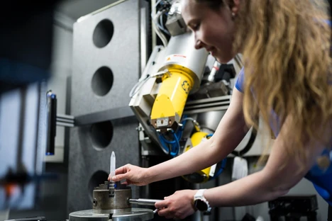
Live Corals Traveled by Train from Vienna to Brno. Scientists Uncovered Their Secret Life
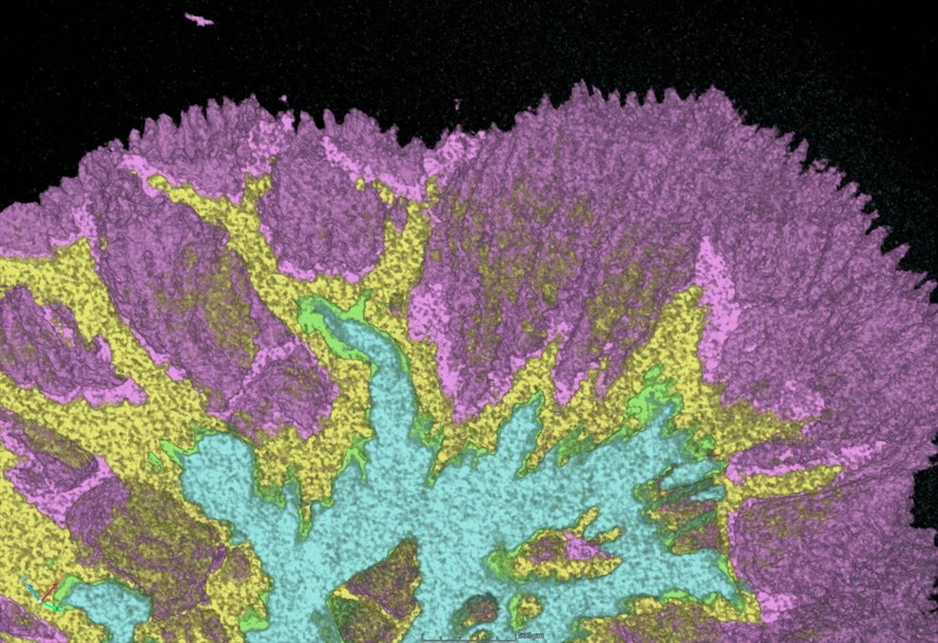
CEITEC BUT: Live Corals Traveled by Train from Vienna to Brno. Scientists Uncovered Their Secret Life
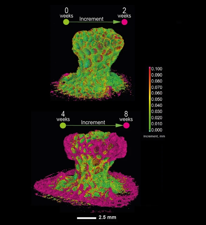
Scientists from CEITEC Brno University of Technology led by Markéta Kaiser have achieved a globally unique breakthrough in coral research. Using the live microCT method, they were able, for the first time ever, to observe the growth of living marine corals in real time and reveal the processes behind the formation of their calcium carbonate skeletons, which had previously remained hidden beneath the surface. The results of the international study, published in the prestigious journal Science Advances, show how corals grow, regenerate, and respond to stress and competing organisms.
The study was carried out through a collaboration between researchers from Brno, Vienna, and several other international institutions. A key role was played by the X-ray Micro and Nano Computed Tomography Laboratory (CTLAB) at CEITEC BUT, where all the crucial data and three-dimensional coral models were produced. CEITEC also shares both the first and corresponding authorship of the study.
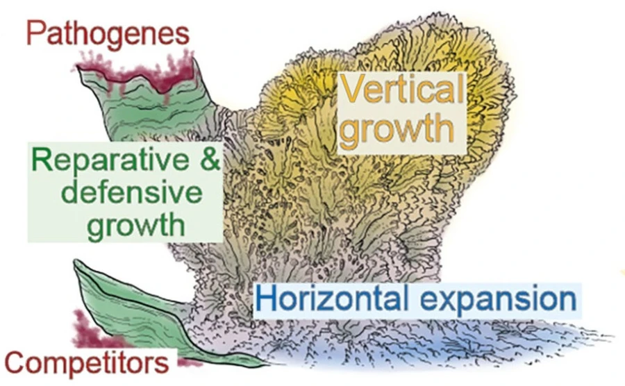
“Thanks to live microCT, we were able, for the first time, to observe what happens beneath the surface of a coral skeleton,” explains Markéta Kaiser from CTLAB at CEITEC BUT. “For example, we witnessed situations where a coral, after being attacked by algae, completely halted its normal growth and began building a protective wall around the threatened area. Once the danger had passed, it was able to resume its original growth pattern.”
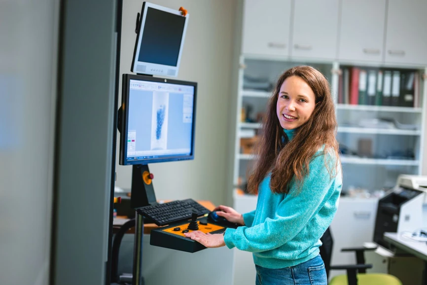 CEITEC BUT/Jakub Rozboud: Live Corals Traveled by Train from Vienna to Brno. Scientists Uncovered Their Secret Life: Markéta Kaiser, CTLAB CEITEC at CEITEC BUT.
The scientists successfully captured coral growth both vertically and horizontally, the formation of protective barriers against competing organisms, and regeneration mechanisms following damage.
The research was exceptionally demanding from a technological standpoint. The team had to develop a special system that enabled living corals to be safely scanned directly in seawater. The coral colonies regularly traveled by train from Vienna to Brno for measurements and, after several hours, were transported back to professional aquarium systems in Vienna.
“At first, it seemed like a somewhat crazy idea. No one knew whether living corals could withstand repeated scanning,” says Kaiser. “Gradually, however, we found that they survived, continued to grow, and provided us with unique data.”
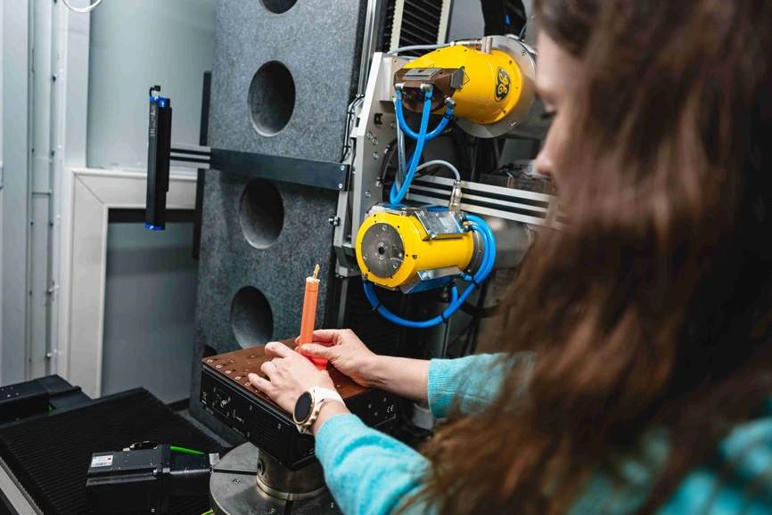 CEITEC BUT/Jakub Rozboud: Live Corals Traveled by Train from Vienna to Brno. Scientists Uncovered Their Secret Life: Markéta Kaiser is placing a coral sample into CT for scanning.
The original concept for the project came from developmental biologist Igor Adameyko of the Medical University of Vienna, who proposed combining marine biology with advanced imaging technologies more commonly used in engineering and technical fields.
CEITEC BUT/Markéta Kaiser: Live Corals Traveled by Train from Vienna to Brno. Scientists Uncovered Their Secret Life: Corals CT - 1.
The findings of the Brno researchers have already attracted the attention of experts in Okinawa, Japan, who specialize in coral reef research. They plan to use the live microCT method to study the effects of light, photosynthesis, and environmental changes on coral growth.
 CEITEC BUT/Markéta Kaiser: Live Corals Traveled by Train from Vienna to Brno. Scientists Uncovered Their Secret Life:Corals CT - 2.
According to the researchers, the project demonstrates the importance of interdisciplinary collaboration between engineering and biological sciences. The research could contribute not only to the study and conservation of coral reefs, but also to fields such as paleontology and materials science.
 CEITEC BUT/Markéta Kaiser: Live Corals Traveled by Train from Vienna to Brno. Scientists Uncovered Their Secret Life: Corals CT - 3.
Original Article
Coral growth, retraction, defense, and regenerative strategies revealed by live microCT
Karina Araslanova, Marketa Kaiser, Anton Fetisov, Diana Gavrik, Tomas Zikmund, Daniel Abed-Navandi, Karel Katovský, Jozef Kaiser, Igor Adameyko
Sci. Adv., 2026, 12, eaee3183
https://doi.org/10.1126/sciadv.aee3183
licenced under CC-BY 4.0
Abstract
Understanding how corals build and remodel their skeletons is key to explaining reef resilience, yet most insights come from static imaging. Using longitudinal live microCT, we tracked the same coral colonies over weeks to months at micrometer resolution. Coral skeleton formation is not a uniform accretion process but a dynamic integration of multiple programs, including vertical and horizontal patterned growth, previously undescribed defensive wall-building against competitors, exploratory edge behavior with reversible expansions and retractions, and skeletal regeneration favoring rapid, imprecise yet effective matrix expansion. Time-resolved imaging links colony-scale growth to microscale events, showing that all modes depend on balances between fusion of skeletal microparticles and layered matrix deposition, guided by tissue prepatterning. Beyond corals, this framework generalizes to studying skeletal dynamics across diverse biomineralizing organisms.